Internship at the Department of Biochemical Engineering & Biotechnology, IIT Delhi
During the summer of 2025, I pursued an internship under Dr Preeti Srivastava at her molecular biology lab in the Department of Biochemical Engineering & Biotechnology, at the Indian Institute of Technology Delhi.
As part of this internship, I conducted a project where I screened soil samples for biosurfactant producing bacteria. This involved collecting and processing samples, and a variety of analytical tests to determine relative biosurfactant production.
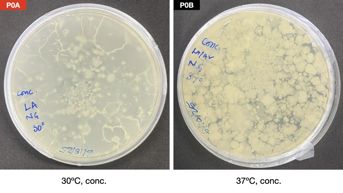
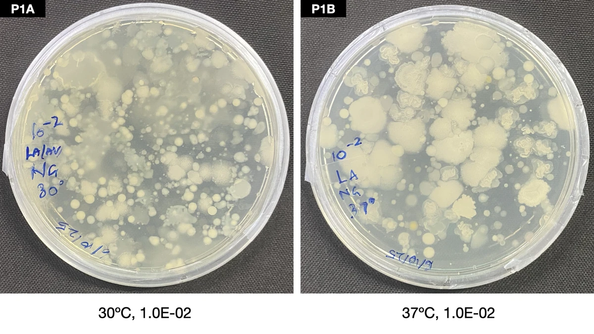
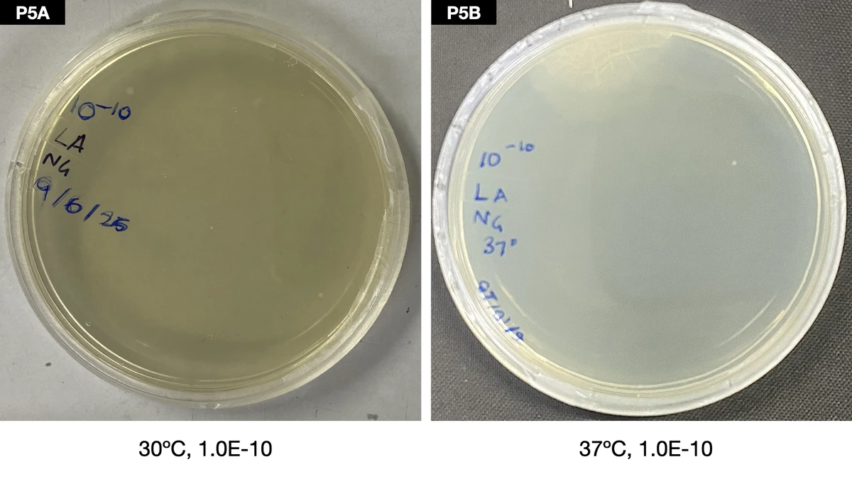
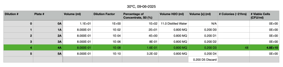
During the first few days, I collected a soil sample (from nearby the department building, nowhere specific) and plated a set five of serial dilutions on Luria Agar plates. Each dilution was spread over two petri plates, and incubated at two temperatures (30ºC and 37ºC) to offer optimum growth temperatures to a variety of possible bacteria in the sample.
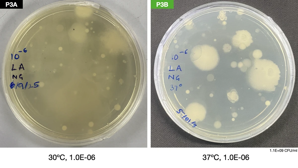
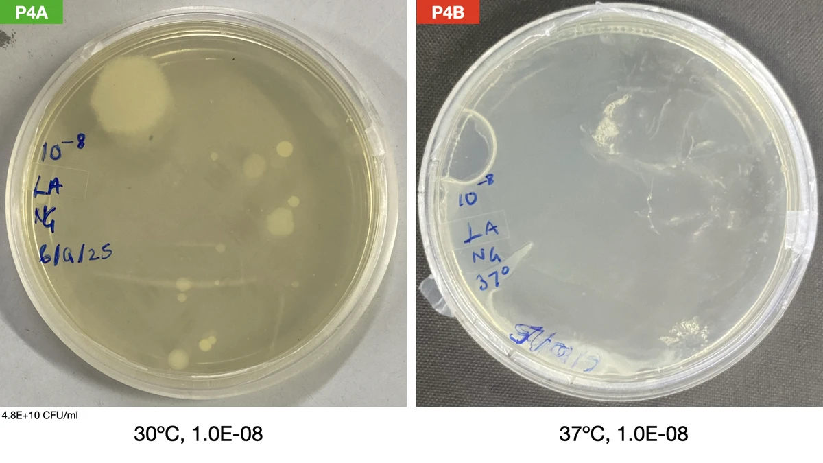
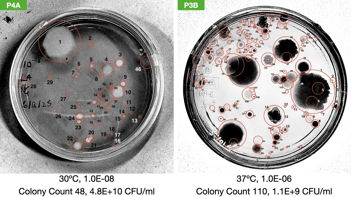
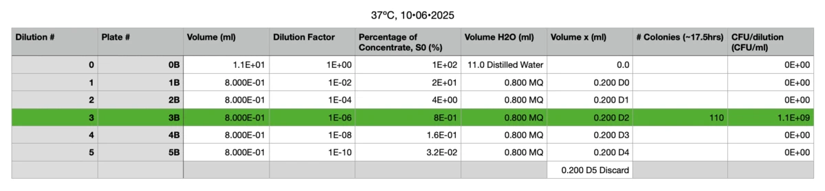
Out of these, P3B and P4A were chosen as best to conduct a CFU count.
It was decided that P3A and P3B showed enough morphological diversity, and had colonies that were separated enough, to plate isolates. Therefore, three individual colonies (with different visible morphologies) each from P3A and P3B were plated on separate Luria Agar plates. Hence, a total of six plates were incubated overnight: three at 30ºC (colonies 1A, 2A & 3A) and three at 37ºC (colonies 1B, 2B and 3B) for biosurfactant testing in the future.
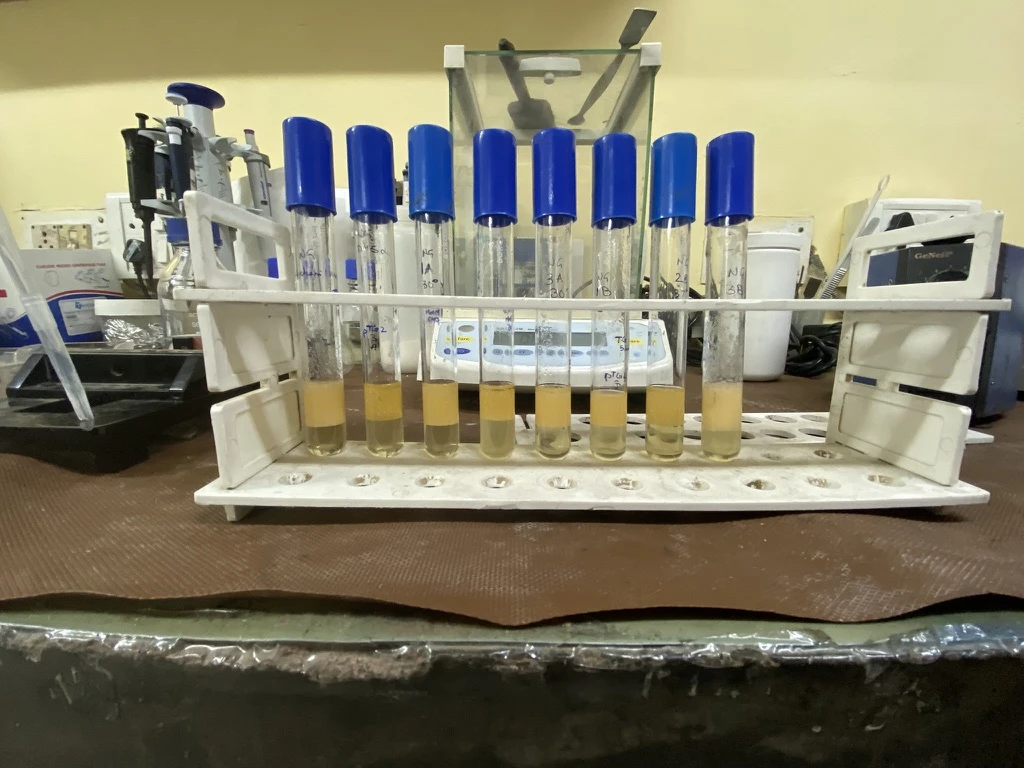
After confirming using the incubated petri plates that each plate contained an isolate, the isolates were incubated in Luria Broth (LB) tubes at their respective temperatures. Positive (B. licheniformis) and negative (the DH5$\alpha$ strain of E. coli) controls for biosurfactant production were also incubated.
The Emulsification Index Assay ($E_{66}\%$, the test was left standing for 66 hours before final measurements were made) was conducted on all eight incubated tubes.
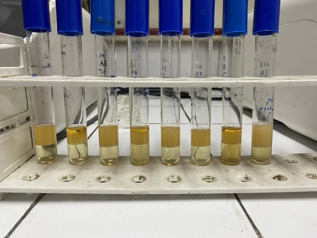
All tubes were centrifuged, and the biosurfactant-containing supernatant was collected. The supernatant was mixed with an equal volume of motor oil, and each tube was agitated using a vortexer for 15 minutes. Each tube was then allowed to stand undisturbed for 66 hours (the standard procedure asks for either 24 or 48 hours, but the experiment was conducted on a Friday and the weekend came in the way), and finally the height of the emulsified layer, and the total liquid, was measured to calculate $E_{66}\%$.
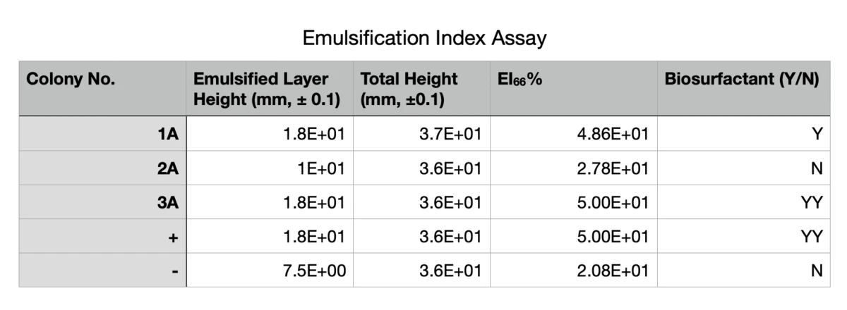
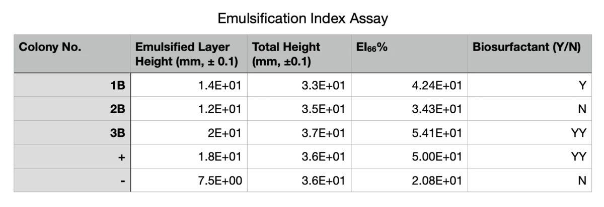
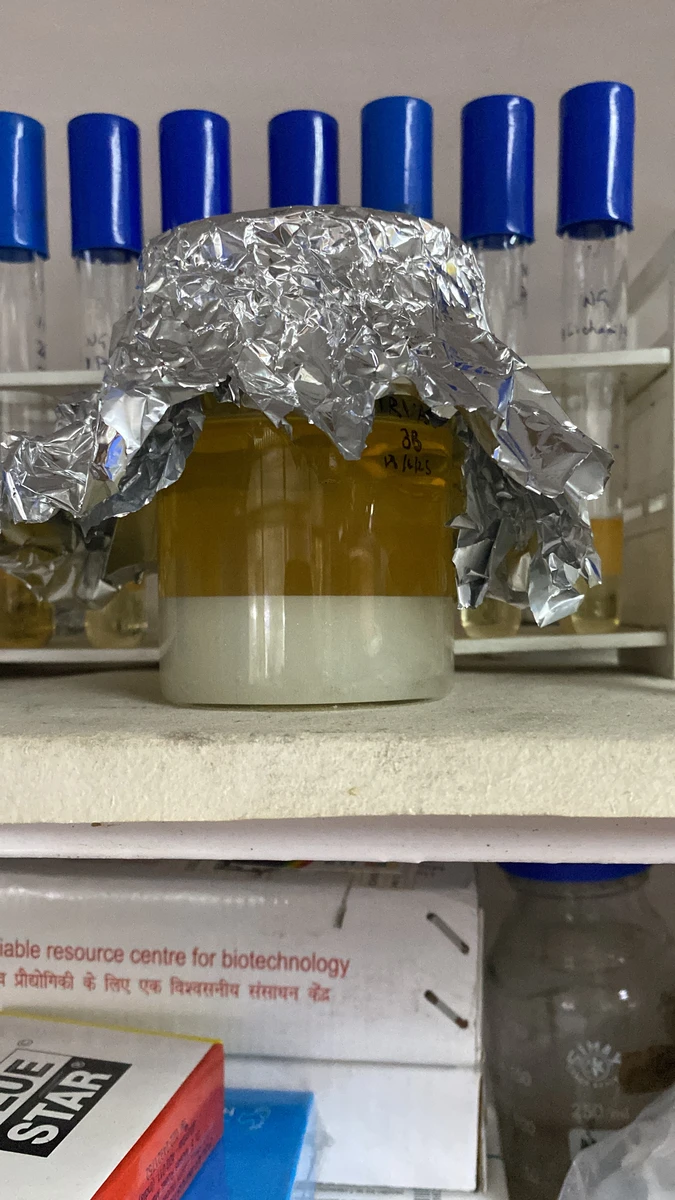
Since sample 3B gave such great results, it was used to inoculate 100ml of Luria Broth for biosurfactant extraction.
The DNA of the bacteria in sample 3B was extracted and purified by cell lysis and silica-column extraction. The 16S gene was amplified using PCR (this is so that the 16S gene can be sent for sequencing, and thus the bacteria in sample 3B can be identified).
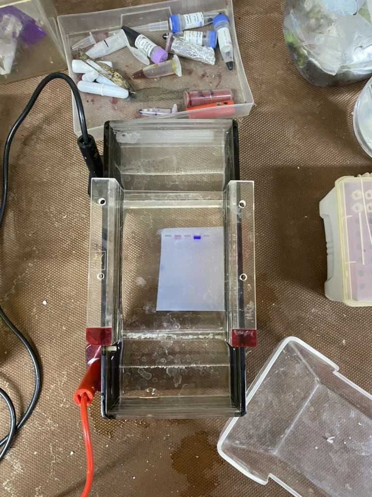
The PCR products were electrophoresed on agarose gel alongside a 1.5kb DNA ladder to verify the PCR worked properly. The verification was successful, and therefore the 16S gene was extracted from the agarose gel.
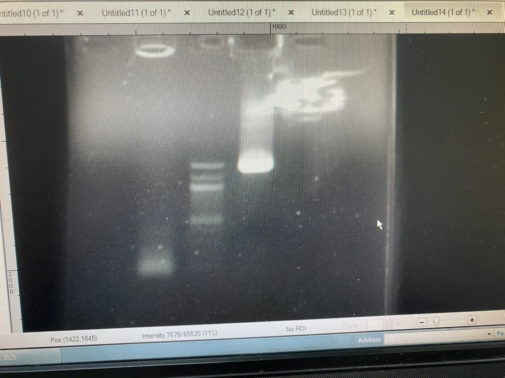
The 16S gene was stored appropriately for sequencing in the future.
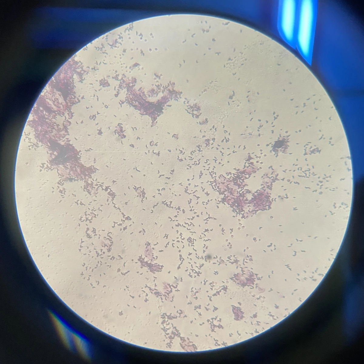
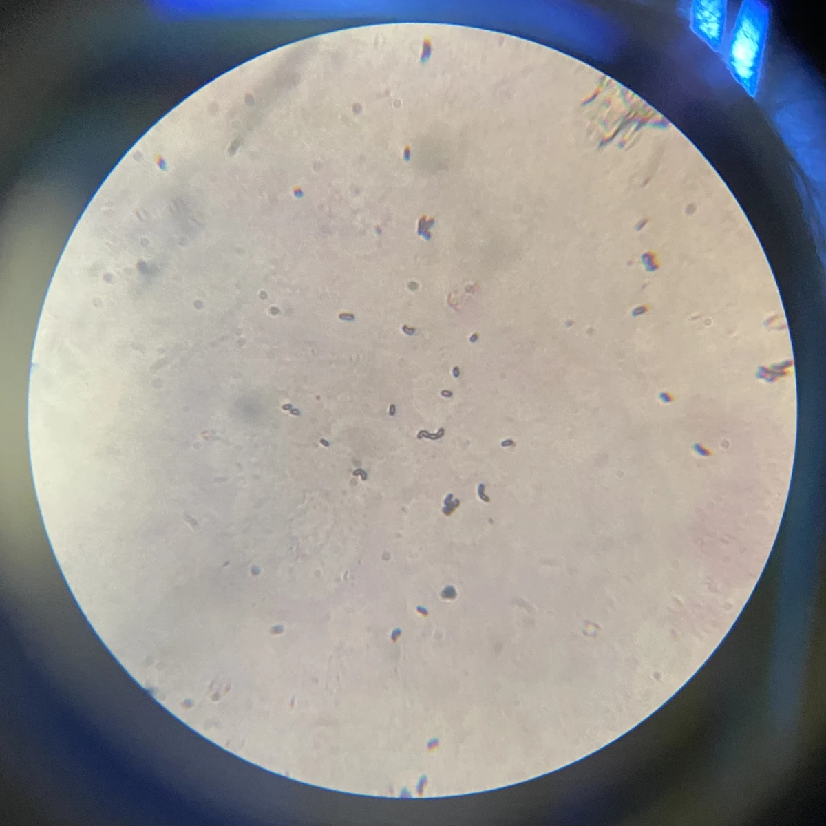
A small amount of culture was also gram-stained. The bacteria were gram-positive.
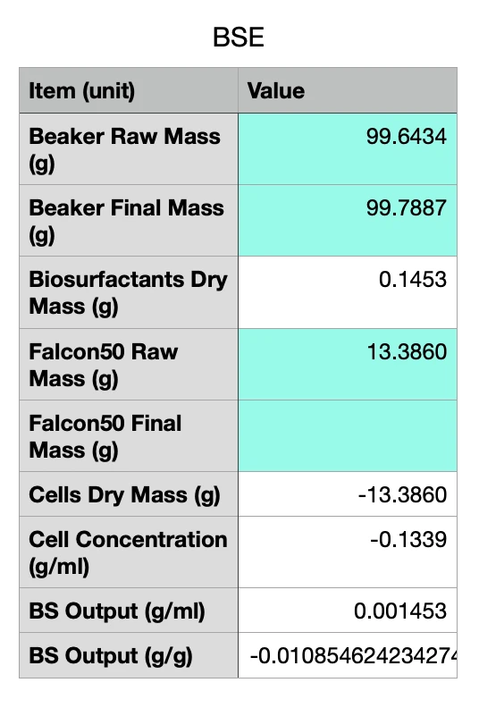
The 100ml culture inoculated with sample 3B was prepared for biosurfactant extraction by centrifuging the culture, collection the supernatant, and adjusting the pH to 2.0. A clean dry beaker, and a clean dry 50ml centrifuge tube (the culture was centrifuged in this tube) were both precisely weighed using an electronic balance. The supernatant was transferred to the weighed beaker, and 100ml of a 2:1 mixture of chloroform-methanol was added to conduct solvent extraction. The whole mixture was magnetically stirred for 45 minutes to ensure complete homogenisation. The beaker was then left to stand for 1.5 hours, after which two distinct layers formed: a white coloured layer at the bottom (chloroform layer, containing the biosurfactant) and a yellow coloured layer on top (methanol layer, containing the LB).
The LB was siphoned off, and the bottom layer was allowed to completely dry in a fume hood. The beaker was then weighed again, which allowed us to calculate the mass of biosurfactant produced by the culture. The centrifuge tube was also allowed to dry. The idea was that this would tell us the the dry mass of the bacterial cells, and then based on this we could estimate the yield of biosurfactant per dry mass of bacterial cells. However, the centrifuge tube was accidentally contaminated, and therefore we couldn’t measure the dry mass, or calculate an accurate estimate of the yield.